The world's first case of OCT in perioperative evaluation of subglottic stenosis in infants
The world's first case of OCT in perioperative evaluation of subglottic stenosis in infants
On the morning of June 3, 2021, a 9-month-old critically ill child was admitted to Guangdong Maternal and Child Health Hospital (Panyu Hospital District). The child had been suffering from recurrent laryngeal crying since birth. Before admission, laryngeal crying worsened with shortness of breath for 3 days and fever for 1 day, manifested as "acute laryngeal obstruction".
CT examination showed subglottic stenosis with fluid dark areas of the mass (Figure 1). To understand the larynx and airway conditions, the patient was examined bronchoscopy and specimens were retained for etiological examination. Under the supervision of PICU, the bronchoscope was entered through the nasopharyngeal cavity, and the nasal secretions were not much, the pharyngeal cavity was not narrow, the epiglottis and bilateral arytenoid cartilage were not softened, the glottis was well opened and closed, and the subglottis showed masses protrusion from the two lateral walls to the lumen, resulting in severe lumen stenosis, and only minimal gaps were left at 12 o 'clock and 6 o 'clock. It was estimated that the bronchoscope with an external diameter of 2.8mm was difficult to pass and might aggravate subglottic edema. The bronchoscope did not pass through the glottis and did not complete the exploration of the lower airway (Figure 2). Diagnosis Conclusion: Subglottic mass, the nature of which needs to be eliminated, further examination is recommended.
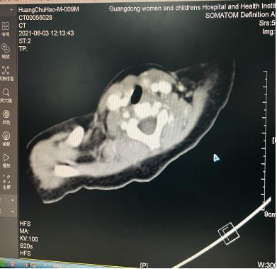
Figure 1. CT image
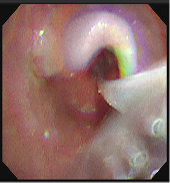
Figure 2 Bronchoscopy
In this case, PICU Director Tang Yuanping immediately thought of the world's leading OCT-respiratory medical imaging technology (Optical coherence tomography equipment, OCT). OCT is a new imaging technology invented by MIT in the 1990s, which has the effect of close to histopathological sections and can carry out in-depth observation and evaluation of airway tissue, so it can be considered as "optical biopsy", opening a new field of medical imaging. It has realized non-invasive, non-radiative, real-time, accurate and stereoscopic visualization of living tissue tomography. Guangzhou Yongshida Medical Technology Co., Ltd. has launched the world's first and only OCT device for human open luminal imaging in 2020, equipped with probes with a diameter of 2.5mm, 1.7mm, 1.0mm and other specifications to adapt to different medical scenarios (Figure 3).
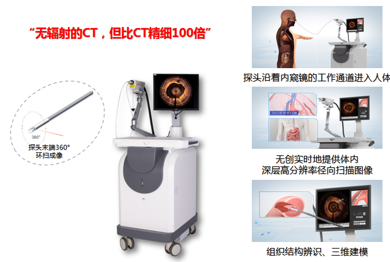
Figure 3 OCTIS respiratory OCT imaging equipment
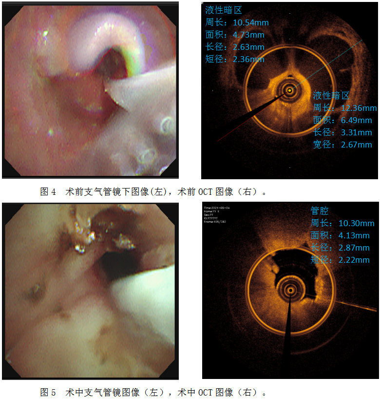
In view of the OCT trial for a period of time in the early stage of PICU and the accumulation of relevant clinical experience, the OCT-guided laser ablation treatment plan was formulated and the operation was carried out on June 4. First, the OCTIS probe was sent into the working channel for observation, and it was found that there were multiple cystic fluid dark areas inside the mass, and the diagnosis was considered to be highly likely to be cysts (Figure 4). Then, the cyst wall is punctured with a sampling needle and the cyst volume is reduced. Finally, holmium laser ablation was performed, and the cyst wall of the tumor was ablated. During the ablation process, OCT was used for several times, and the ablation was stopped after it was confirmed that no cystic structure was found (FIG. 5).
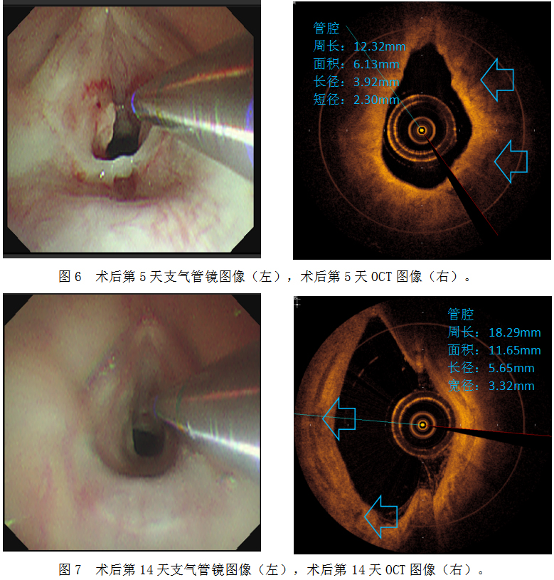
On the 5th day after surgery, OCT was used to observe the airway hierarchy and small holes caused by suspected cyst collapse could be seen (Figure 6). On the 14th day, OCT imaging showed that the subglottic tissue structure was normal, no obvious surgical wounds were observed, and no cysts were observed (FIG. 7).

In this case, when CT, bronchoscopy and other examination methods could not evaluate the nature and extent of the lesion of the subglottic mass, OCT technology was used to observe and evaluate the lesion site and formulate a reasonable surgical plan. OCT was used to observe and evaluate the lesion during operation. In postoperative review, OCT was used to track the evaluation of surgical efficacy and to judge the prognosis. OCT examination covers the perioperative period of children and plays an important role in escorting the treatment of children.
The most narrow part of the airway in infants and young children is under the glottis. After the occurrence of the lesion, the bronchoscope can only examine the mucosal surface, and often cannot reach the lesion site. OCT probe has a small diameter, can pass through the subglottic lesion site, and perform non-invasive, non-radiative, real-time high-definition imaging of the submucosa 2-3mm tissue, which can improve the acceptance of the examination in children and improve the accuracy of assessment and diagnosis. This case is the world's first case of OCT application in perioperative period of laser ablation for subglottic stenosis in infants, which has important clinical reference significance (FIG. 8).
This case also provides new ideas for the development of OCT. OCT can be safely and effectively applied in the preoperative assessment and diagnosis of infant respiratory diseases and guide the formulation of surgical plans. Intraoperative monitoring and evaluation of surgical results; Following up the postoperative review and follow-up of the surgical effect, OCT is of great clinical significance in the diagnosis and treatment of infant subglottic diseases.
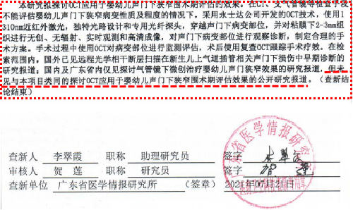
Figure 8 Novelty search report


