Respiratory OCT
Respiratory OCT
OCTIS® is currently the world's only OCT device suitable for airway clinical observation, and is the world's first open luminal optical interference tomography device that has obtained the national medical device registration certificate, which has a world-leading milestone in the development of OCT technology in the clinical application of open luminal devices.
Academician Zhong Nanshan recommended Ostian Respiratory OCTIS® system to Sinopath as "the first in China and at the world's advanced level." It will fill the gap in the current domestic precision imaging medical equipment."
Index | Respiratory tract OCT® |
Resolution | 10-20μm |
Detection range | Airway levels 1-9 |
Observation range | Airway lumen, bronchial mucosa, submucosa, smooth muscle, cartilage, glands |
Clinical conclusion
Respiratory OCTIS affects the identification rate of airway tissue structure by the device to 97.02%, which can dynamically, real-time, accurate, non-invasive, and penetrable observe the fine tissue structure of the airway, solving the problem that it is difficult to obtain near- microscopic images of the human body cavity at present, and greatly improving the efficiency of diagnosis and treatment.
Clinical application scenario
1. Diagnosis of moderate and severe long-term lung diseases
OCT can directly and quantitatively evaluate the pathological classification and airway remodeling of COPD, asthma and conventional lung diseases, and provide grading and quantitative indicators for chronic airway diseases.
2. Identify benign and malignant airway lesions
For patients with pulmonary nodules, shadows or ground glass structures found in routine imaging screening, if it is necessary to perform fiberoptic bronchoscopy for biopsy, OCT can predict the benign and malignant lesions in real time, and achieve the effect of distinguishing early lung cancer and guiding biopsy.
3. Tracheoscopy assisted therapy and surgery
In a variety of surgical and therapeutic scenarios, such as BT (thermal ablation therapy), airway laser therapy, airway stenosis/dilatation, lung volume reduction, and stent implantation, OCT is used to monitor the changes of the whole and local tissues of the airway in real time to improve the accuracy of treatment and the efficiency of diagnosis and treatment.
4. Long-term follow-up of airway diseases
Patients with moderate/severe lung cancer, COPD, asthma, and pneumonia receive regular follow-up examinations after medication or surgery through airway OCT.
5. Multi-disciplinary joint application
Pulmonary parenchyma, peripheral pulmonary or thoracic puncture examination and surgical operation can be assisted by airway OCT to accurately observe focal tissue.
Clinical application cases (corresponding to three pages)
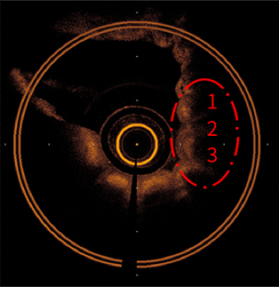
01 OCT imaging features of lung cancer
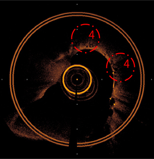
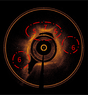
At present, there are risks of missed diagnosis and misdiagnosis in biopsy sampling. Respiratory OCTIS can distinguish benign and malignant local lesions in the airway and common tumor types before biopsy, and achieve non-invasive early screening, which is expected to increase the positive rate of tracheoscopic biopsy to more than 80%.
Squamous carcinoma
1. Scaly bulge
2. Large dark spots
3: Swelling
adenocarcinoma
1. Black spot distribution
Small cell carcinoma
1. The pipe wall is raised
2. Post-epidermal dark spots
3. Lamina propria thickening
02 OCT imaging features of COPD
At present, FEV1 cannot determine the location of lesions. Respiratory OCTIS can observe the structural changes of grade 3-9 bronchus, accurately identify early lesions of small airways, and quantitatively evaluate the degree of airway narrowing and wall thickening, which can assist in COPD classification.
OCT image of airway cross-section
Compared with non-smokers, the mucosa of COPD patients was thickened significantly and the submucosa was blurred.
(Three pictures in total)


OCT image of longitudinal section of airway
Compared with non-smokers and heavy smokers with normal lung function, the airway lumen is significantly narrowed in COPD patients.
(Three pictures in total)


Source of information:
Ding M , Chen Y , Guan W J , et al. Measuring Airway Remodeling in Patients with Different COPD Staging Using Endobronchial Optical Coherence Tomography[J]. Chest. 2016 Dec; 150(6): 1281-1290.
03 OCT image characteristics of asthma
Respiratory OCTIS evaluated the severity of asthma and the effect of BT therapy by examining changes in the number of airway folds, smooth muscle and airway basal layer in OCT images.
(Illustration: There are more folds in the airway)
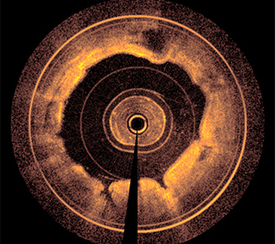
(Illustration: smooth muscle hyperplasia and tube wall thickening)
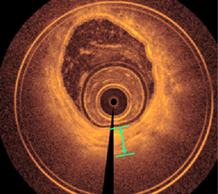
(Illustration: Thickening of airway mucosa epithelium)
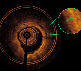
Source: Yongstar Ostin OCTIS® Respiratory OCT clinical study data


